Advertisement
Advertisement
Members of the team of heart disease specialists who performed the ground-breaking operation. Photo: Shirley Zhao

How a 3D-printed heart helped surgeons pull off world-first operation on Hong Kong woman

Team at Queen Elizabeth Hospital replaces two heart valves through woman’s blood vessels in a single operation in what is a world’s first
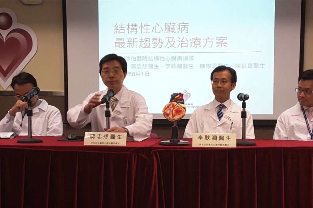
Specialists at Queen Elizabeth Hospital have successfully conducted the world’s first ever heart surgery involving the replacement of two valves through blood vessels in a single operation.

Doctors conducted the highly complex operation on a 77-year-old woman.

The eight-person team used 3D-printing technology to create a detailed model of the patient’s heart to allow better planning and more practice, thus ensuring more precision during the actual procedure, which was carried out over four hours on June 27.

The team, which specialises in minimally invasive surgery on patients with heart disease from birth or developing with wear and tear, said traditional open heart surgery required cutting open the chest, leading to greater risk. This procedure is also not preferred for older or weaker patients as recovery can be lengthy.

The specialists said a better option was performing heart surgery through blood vessels, which requires only opening a hole in the body to access the targeted area. The procedure can minimise harm to a patient’s body and shorten the recovery time.

The patient, identified only by her surname Shum, had two damaged heart valves – the mitral valve on the left side and the tricuspid valve on the right – caused by inflammation. This led to blood in the heart flowing backwards, which can result in heart failure.

Shum has undergone three open heart operations since 1973 and has had two damaged valves replaced by artificial ones.

But her condition worsened early this year with symptoms of heart failure. An ultrasound scan of her heart showed medium to severe narrowing of the aortic valve on the left side and severe narrowing of the artificial tricuspid valve.

“Given Shum’s age and her condition, it would be highly risky to perform another open heart surgical operation,” said Dr Michael Lee Kang-yin, a consultant cardiologist at the hospital and a member of the specialist team. “Replacing the valves through the blood vessel was the only suitable plan.”

The procedure was to insert a new artificial valve, made from a cow’s heart tissue on a metal frame, into her old, broken artificial tricuspid valve through a 5-6mm opening in a main vein on her right upper thigh. This was done by leading an 80-100mm guide wire through the vein all the way to the heart.

The operation went smoothly thanks to the preparation and practice on the 3D printed heart model. Inserting the replacement valve took about three hours, but doctors then found the existing aortic valve was narrower than expected. They therefore decided to replace the valve with an artificial one made of pig’s heart tissue in the same operation.

Surgery started at 9am and ended at about 1pm.

Because of the nature of the surgery, Shum was able to sit and walk one day after the operation and was released from hospital a week later. If she had undergone open heart surgery, she could have suffered complications such as stroke or inflammation and could have been in hospital much longer, team member and consultant cardiologist Dr Sammy Chiang Chung-seung said.

Chiang said the hospital’s structural heart disease division, which has 15 specialists, obtained extra funding from the Hospital Authority to hire two more staff, including a doctor and a nurse.

With the extra manpower, the division planned to increase the number of minimally invasive operations from the current 20-30 a year to about 80 or more, he said.

“Hopefully we can start developing and expanding our services from this year,” Chiang said.

MEDICAL MILESTONES

Patient: female, 77 years old with three previous open heart operations

Hospital: Queen Elizabeth

Length of surgery: four hours

Team: one radiologist, one cardiac anaesthetist, one cardio-surgeon, five cardiologists

Valves involved: tricuspid valve (right) and aortic valve (left)

Materials used in the artificial valves: cow’s or pig’s heart tissue mounted on top of a metal frame

Opening: 5-6mm on the right femoral vein from the upper thigh

WORLD BEATERS

World first medical developments in Hong Kong:

October 1996: Specialists from Queen Mary Hospital transplanted the right half of a liver from a living adult to another.

October 2014: Queen Mary Hospital transplanted a liver, which had already been transplanted once 11 years earlier to a Hepatitis B sufferer, to another patient with the same condition. The surgery took 11 hours.

July 2015: Queen Mary Hospital formed liver parts taken from two Macau sisters into one liver and transplanted it to their father, who was suffering from liver failure. The operations involving the sisters and the father happened simultaneously, taking 55 minutes.

March 2016: Researchers from the University of Hong Kong and Polytechnic University announced that they had developed the world’s first robot capable of performing abdominal surgery without cutting into a patient’s body. The new system allows small robotic parts to be placed inside a patient via a tube inserted into natural openings. The parts can then be assembled inside the body.

This article appeared in the South China Morning Post print edition as: Groundbreaking heart op performed on elderly woman
Post